
.svg)


Clinical Vision - Platform
AI-assisted MRI review, from lesion detection to signed clinical report.
Confidential Medical Company
About The Project
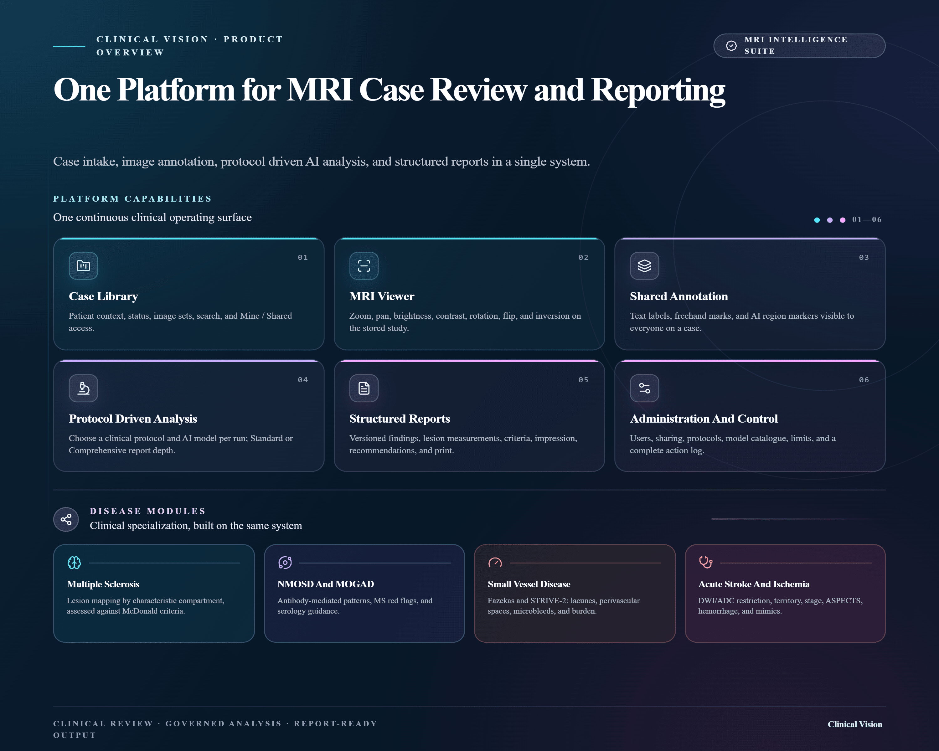
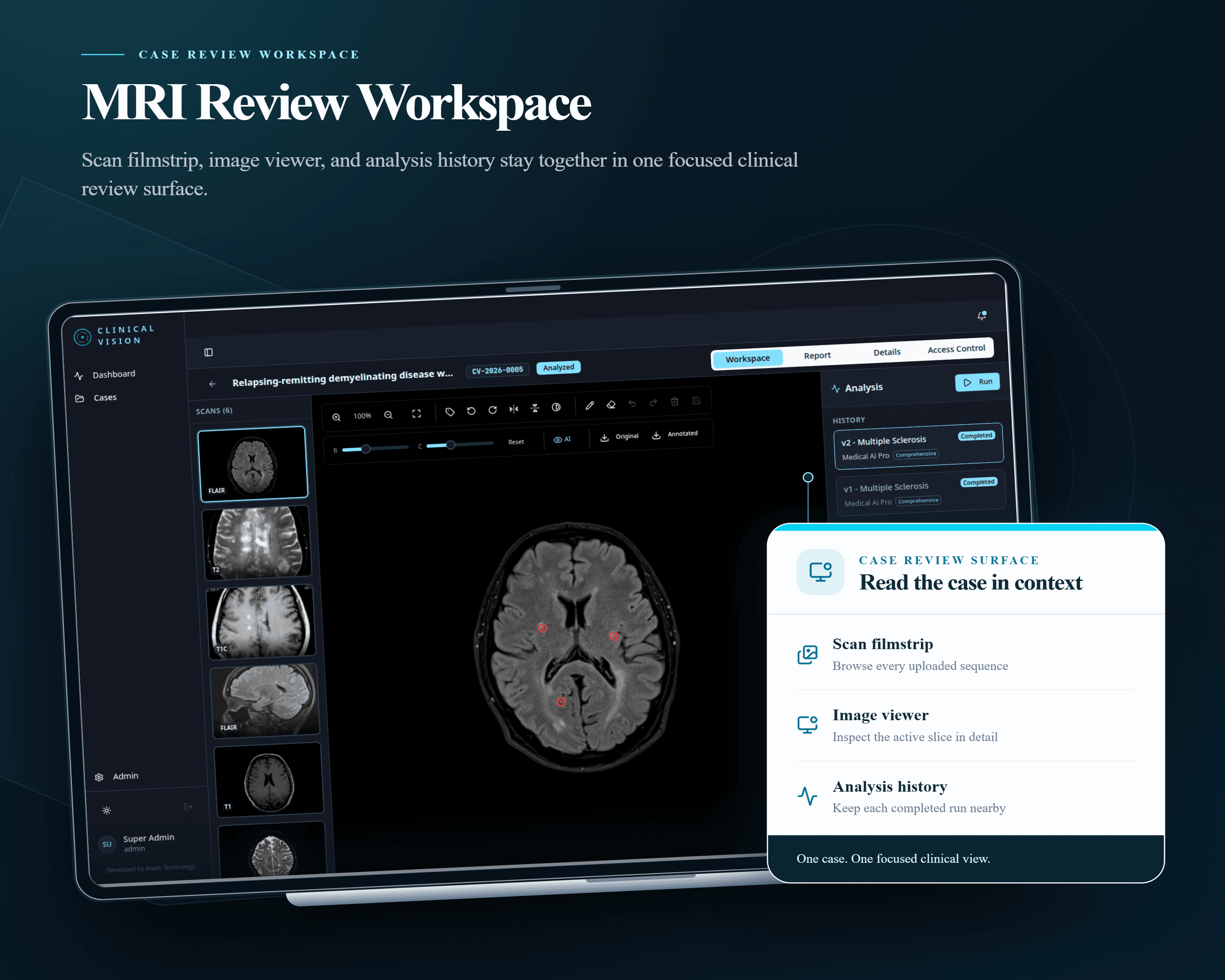
Clinical Vision is an AI platform for MRI review, built by Areeb Innovative Technologies. The core is a trained multi-slice detection model. Standard 2D models read one slice at a time; this one reads consecutive slices together, so it reasons about lesion continuity across the volume the way a specialist does. It outputs candidate lesion regions with confidence scores, mapped directly onto the scan. A second AI layer turns detection into documentation: a chosen clinical protocol runs against a chosen model to produce a structured report with findings, criteria scoring, lesion measurements, and an impression. The output is a draft for clinician review and sign off, not an autonomous diagnosis. Disease modules cover multiple sclerosis (McDonald), NMOSD and MOGAD, small vessel disease (Fazekas, STRIVE-2), and acute stroke (ASPECTS). Arabic and English, light and dark.
Key Challenges
-
Brain MRI varies widely in contrast, resolution, orientation, and noise by scanner and protocol, so the model needed preprocessing and normalization pipelines plus curated multi-center data to hold up outside its training distribution.
-
Lesions are small, faint, and easy to confuse with normal anatomy on a single slice, which is why detection reads slices in sequence rather than independently.
-
Pixel-level annotated MRI is scarce, so augmentation, semi-supervised learning, and cross-dataset validation carried performance beyond what the labeled set alone supported.
-
Over-detection creates false alarms and under-detection misses real lesions, so the operating point was set through custom loss functions and evaluation rounds with neurologists.
-
A confidence number alone does not earn clinical trust, so every flag had to become visible evidence on the scan tied to a criteria score in the report.
-
Different model providers return different response shapes, and each has to land in the same structured report format.
-
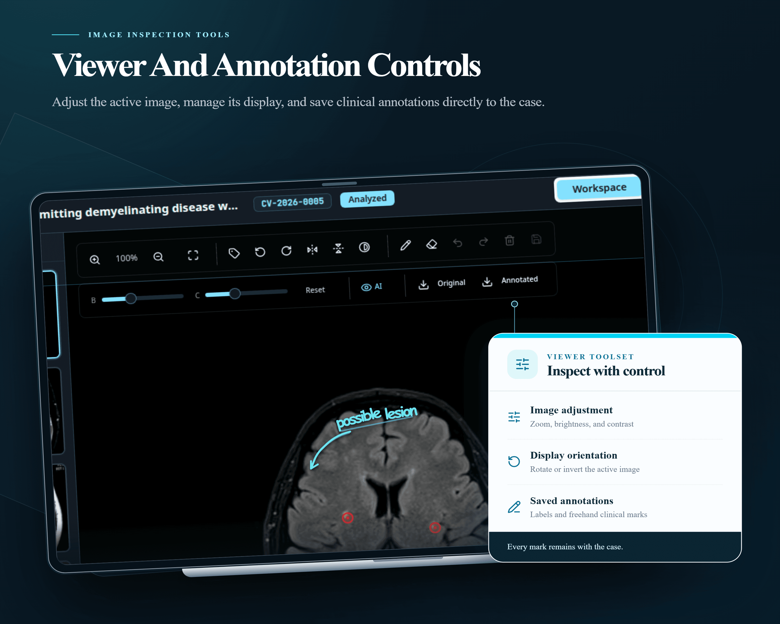
Overlays are stored as normalized coordinates, so every rotation, flip, or inversion has to remap them or the markings drift off the anatomy.
-
Analysis is long-running and can fail partway, so runs need visible state and per-run versioning.
-
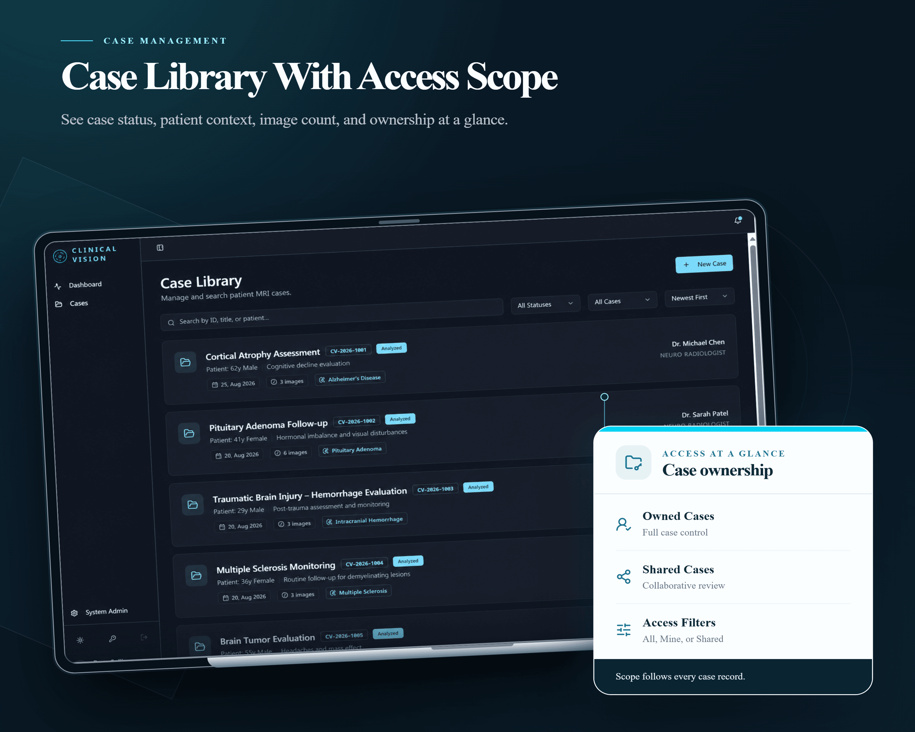
Access is scoped per case, not per account, and patient images stay authenticated and encrypted through upload, storage, and export.
What we delivered
- A trained multi-slice detection model producing lesion regions with confidence scoring, surfaced as overlays in the viewer.
- An analysis engine that runs a selected protocol against a selected model, tracks run state and cost, and writes a versioned structured report with findings, marked regions, criteria scoring, and an impression.
- A provider-agnostic model layer with a configurable catalog, normalizing every provider response into one report schema.
- GPU-optimized inference and streamlined preprocessing for turnaround in minutes.
- A browser MRI viewer with zoom, pan, brightness and contrast, rotation, flip and inversion, shared labels, freehand pen with undo and redo, and layered overlay visibility.
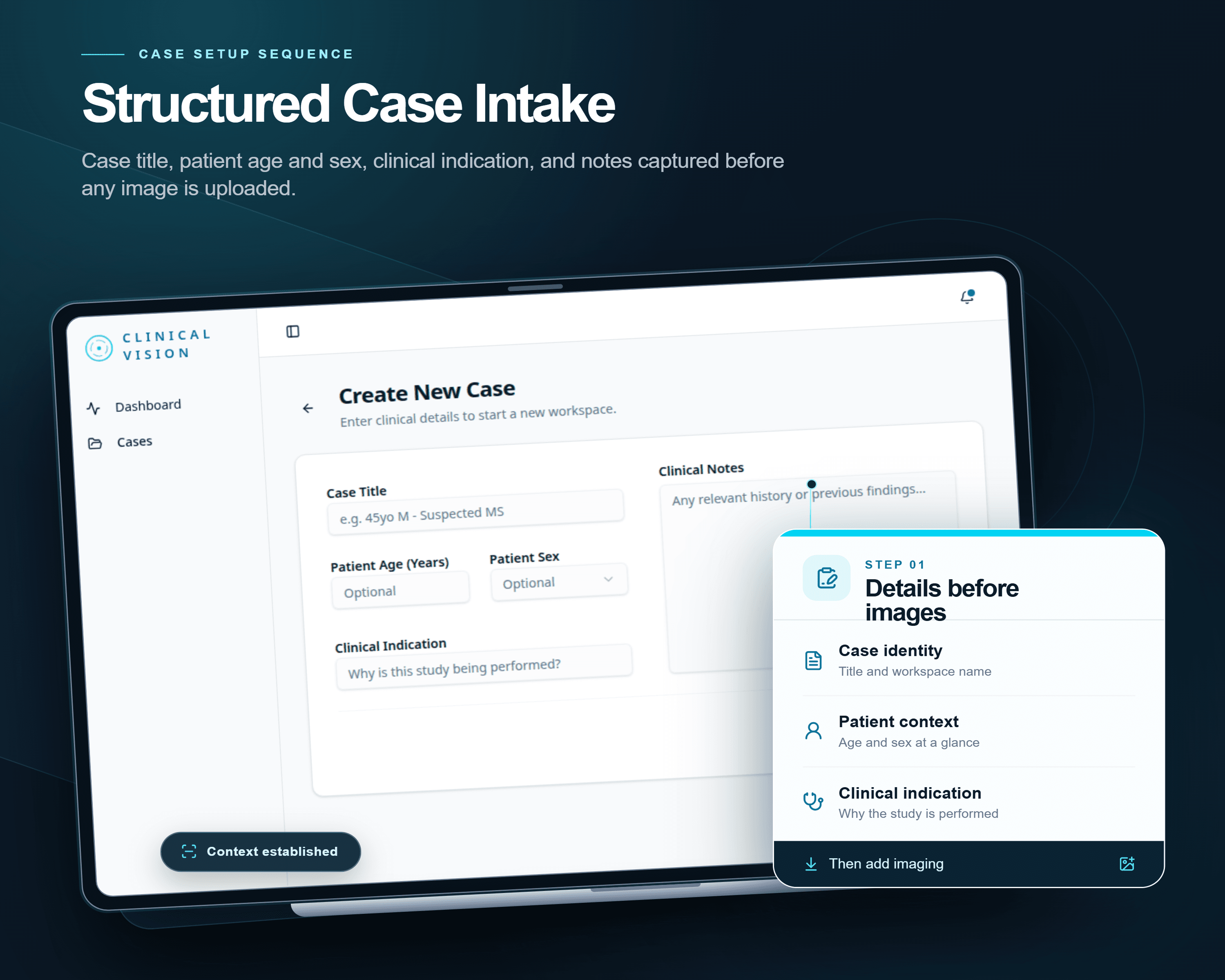
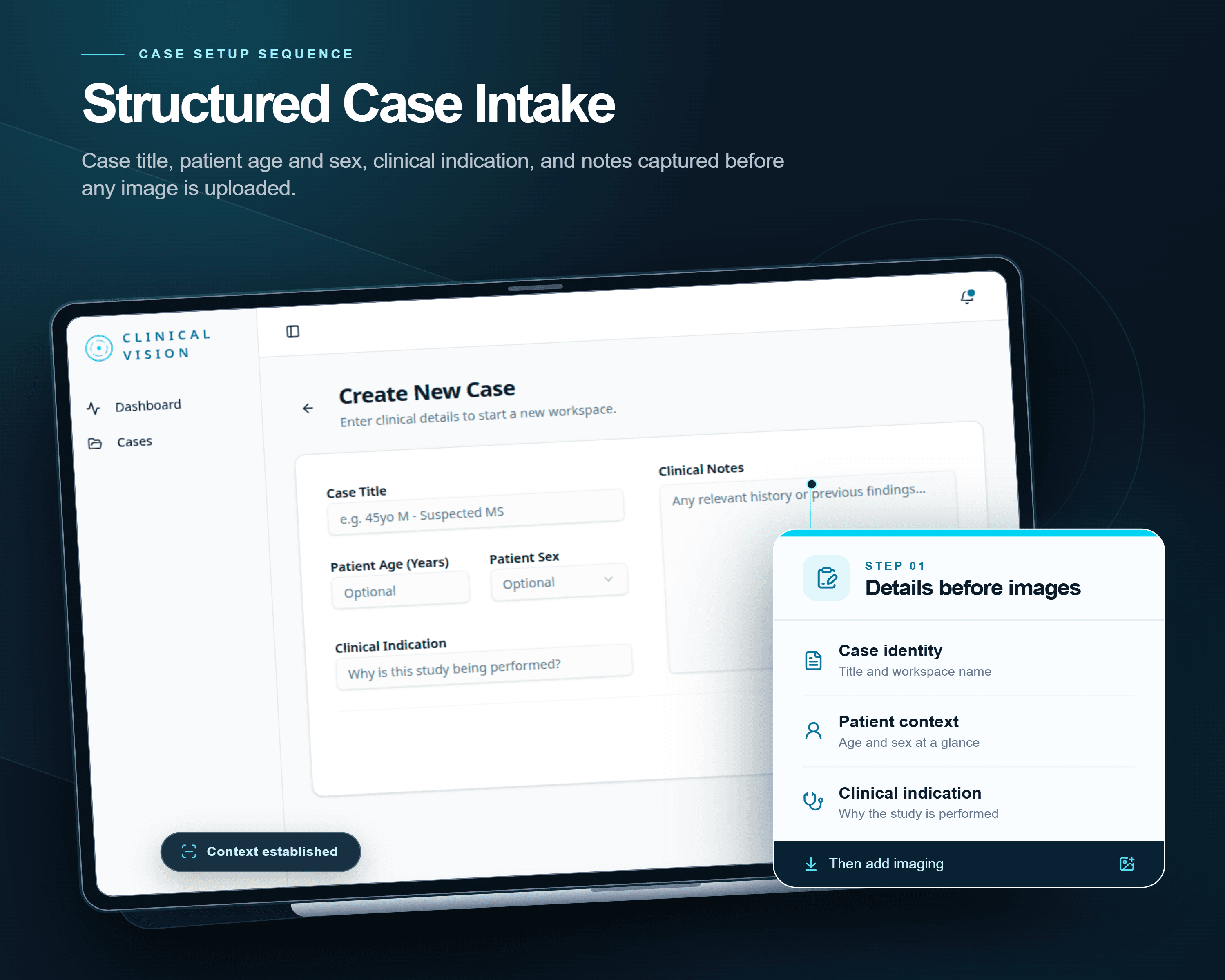
- A case library with search, status and ownership filters, patient context, and share-based access control.
- A printable report covering study metadata, clinical context, technique, findings, lesion measurements, criteria, impression, recommendations, and sign off, plus image export.
- An admin area for users, protocols, model catalog and pricing, usage limits, and a full activity log.
Outcome
Detection, review, and reporting sit in one system, so a case never leaves the platform between steps. Every AI flag is visible evidence on the scan rather than an unexplained result, which is what makes it usable in a clinical decision. Shared annotations mean every reviewer works on the same study. Protocols and models are configuration, so an organization changes what the system analyzes, and what it costs, without a code change. Every action is recorded against the user who made it.